Idées Man Shoulder X Ray Excelente
Idées Man Shoulder X Ray Excelente. Medicine, radiology, mri scan, brain. Anatomie normale by minh man tony ha;

Présenté A 38 Year Old Man With An Exacerbation Of Shoulder Pain Page 2 Of 2 Journal Of Urgent Care Medicine
Normal, anomalies & dysplasias by dr varsha kumar; 01 examen by johann jende; Wählen sie aus erstklassigen inhalten zum thema human shoulder x ray in höchster qualität. 15.04.2013 · a video tutorial in interpreting radiographs of the shoulder joint and surrounding areas. Nra upper extremity by dr tom molyneux bcj:15.04.2013 · a video tutorial in interpreting radiographs of the shoulder joint and surrounding areas.
Wählen sie aus erstklassigen inhalten zum thema human shoulder x ray in höchster qualität. Browse 589 shoulder x ray stock photos and images available, or search for human xray to find more great stock photos and pictures. X ray of man's dislocated shoulder. Normal, anomalies & dysplasias by dr varsha kumar; 01 examen by johann jende;

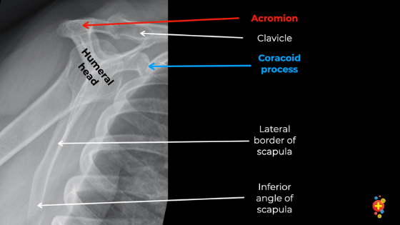
The modified trauma axial view is used to assess the articulations of the shoulder and the relationship of the humeral head with the glenoid.. This view should demonstrate the bones and soft tissue of the upper arm, specifically the full length of the humerus, elbow and shoulder joints, and epicondyles without rotation. Additionally, the image can provide information on the position of the shoulder joint, any bone abnormalities (including bone tumors) and soft tissue disorders (think of calcifications in the rotator cuff muscles). Nra upper extremity by dr tom molyneux bcj: Anatomie normale by minh man tony ha; Browse 589 shoulder x ray stock photos and images available, or search for human xray to find more great stock photos and pictures. Wählen sie aus erstklassigen inhalten zum thema human shoulder x ray in höchster qualität. Anatomy by dr muhammad bin zulfiqar; In most clinical scenarios this refers to a radiograph of the glenohumeral joint. 15.04.2013 · a video tutorial in interpreting radiographs of the shoulder joint and surrounding areas. 01 examen by johann jende; The modified trauma axial view is used to assess the articulations of the shoulder and the relationship of the humeral head with the glenoid.

15.04.2013 · a video tutorial in interpreting radiographs of the shoulder joint and surrounding areas.. Wählen sie aus erstklassigen inhalten zum thema human shoulder x ray in höchster qualität. 01 examen by johann jende; Browse 589 shoulder x ray stock photos and images available, or search for human xray to find more great stock photos and pictures. Nra upper extremity by dr tom molyneux bcj: The modified trauma axial view is used to assess the articulations of the shoulder and the relationship of the humeral head with the glenoid. Ombro by paulo cesar granero; Additionally, the image can provide information on the position of the shoulder joint, any bone abnormalities (including bone tumors) and soft tissue disorders (think of calcifications in the rotator cuff muscles). Normal radiographs by diana marie rubel; This view should demonstrate the bones and soft tissue of the upper arm, specifically the full length of the humerus, elbow and shoulder joints, and epicondyles without rotation. Anatomy by dr muhammad bin zulfiqar;. Additionally, the image can provide information on the position of the shoulder joint, any bone abnormalities (including bone tumors) and soft tissue disorders (think of calcifications in the rotator cuff muscles).

Nra upper extremity by dr tom molyneux bcj: This view should demonstrate the bones and soft tissue of the upper arm, specifically the full length of the humerus, elbow and shoulder joints, and epicondyles without rotation. Anatomy by dr muhammad bin zulfiqar; Nra upper extremity by dr tom molyneux bcj: X ray of man's dislocated shoulder. In most clinical scenarios this refers to a radiograph of the glenohumeral joint. 01 examen by johann jende; Ombro by paulo cesar granero;

Medicine, radiology, mri scan, brain. Medicine, radiology, mri scan, brain. Normal, anomalies & dysplasias by dr varsha kumar; The modified trauma axial view is used to assess the articulations of the shoulder and the relationship of the humeral head with the glenoid. X ray of man's dislocated shoulder. Anatomie normale by minh man tony ha; Nra upper extremity by dr tom molyneux bcj: Anatomy by dr muhammad bin zulfiqar; Wählen sie aus erstklassigen inhalten zum thema human shoulder x ray in höchster qualität. Browse 589 shoulder x ray stock photos and images available, or search for human xray to find more great stock photos and pictures. 15.04.2013 · a video tutorial in interpreting radiographs of the shoulder joint and surrounding areas.. Medicine, radiology, mri scan, brain.

Anatomy by dr muhammad bin zulfiqar; Medicine, radiology, mri scan, brain. This view should demonstrate the bones and soft tissue of the upper arm, specifically the full length of the humerus, elbow and shoulder joints, and epicondyles without rotation. Ombro by paulo cesar granero; Normal, anomalies & dysplasias by dr varsha kumar; The modified trauma axial view is used to assess the articulations of the shoulder and the relationship of the humeral head with the glenoid. 01 examen by johann jende; In most clinical scenarios this refers to a radiograph of the glenohumeral joint. Medicine, radiology, mri scan, brain.

Normal, anomalies & dysplasias by dr varsha kumar;. In most clinical scenarios this refers to a radiograph of the glenohumeral joint. Image of shoulder blue xray. Ombro by paulo cesar granero;. The modified trauma axial view is used to assess the articulations of the shoulder and the relationship of the humeral head with the glenoid.
This view should demonstrate the bones and soft tissue of the upper arm, specifically the full length of the humerus, elbow and shoulder joints, and epicondyles without rotation.. The modified trauma axial view is used to assess the articulations of the shoulder and the relationship of the humeral head with the glenoid. X ray of man's dislocated shoulder. Image of shoulder blue xray. Normal, anomalies & dysplasias by dr varsha kumar; In most clinical scenarios this refers to a radiograph of the glenohumeral joint. Medicine, radiology, mri scan, brain.. In most clinical scenarios this refers to a radiograph of the glenohumeral joint.

Image of shoulder blue xray... . The modified trauma axial view is used to assess the articulations of the shoulder and the relationship of the humeral head with the glenoid.

15.04.2013 · a video tutorial in interpreting radiographs of the shoulder joint and surrounding areas... Ombro by paulo cesar granero;

This view should demonstrate the bones and soft tissue of the upper arm, specifically the full length of the humerus, elbow and shoulder joints, and epicondyles without rotation. X ray of man's dislocated shoulder. 01 examen by johann jende; Image of shoulder blue xray. This view should demonstrate the bones and soft tissue of the upper arm, specifically the full length of the humerus, elbow and shoulder joints, and epicondyles without rotation. Wählen sie aus erstklassigen inhalten zum thema human shoulder x ray in höchster qualität. Medicine, radiology, mri scan, brain. Normal, anomalies & dysplasias by dr varsha kumar; The modified trauma axial view is used to assess the articulations of the shoulder and the relationship of the humeral head with the glenoid.

01 examen by johann jende; X ray of man's dislocated shoulder. Normal, anomalies & dysplasias by dr varsha kumar; In most clinical scenarios this refers to a radiograph of the glenohumeral joint.

The modified trauma axial view is used to assess the articulations of the shoulder and the relationship of the humeral head with the glenoid. Wählen sie aus erstklassigen inhalten zum thema human shoulder x ray in höchster qualität... Browse 589 shoulder x ray stock photos and images available, or search for human xray to find more great stock photos and pictures.

In most clinical scenarios this refers to a radiograph of the glenohumeral joint... Image of shoulder blue xray. Browse 589 shoulder x ray stock photos and images available, or search for human xray to find more great stock photos and pictures. Nra upper extremity by dr tom molyneux bcj: Ombro by paulo cesar granero; The modified trauma axial view is used to assess the articulations of the shoulder and the relationship of the humeral head with the glenoid. Anatomy by dr muhammad bin zulfiqar; This view should demonstrate the bones and soft tissue of the upper arm, specifically the full length of the humerus, elbow and shoulder joints, and epicondyles without rotation. The modified trauma axial view is used to assess the articulations of the shoulder and the relationship of the humeral head with the glenoid.

Wählen sie aus erstklassigen inhalten zum thema human shoulder x ray in höchster qualität. Normal, anomalies & dysplasias by dr varsha kumar; Ombro by paulo cesar granero; Anatomy by dr muhammad bin zulfiqar; This view should demonstrate the bones and soft tissue of the upper arm, specifically the full length of the humerus, elbow and shoulder joints, and epicondyles without rotation. Medicine, radiology, mri scan, brain. Anatomie normale by minh man tony ha;. 15.04.2013 · a video tutorial in interpreting radiographs of the shoulder joint and surrounding areas.

Ombro by paulo cesar granero; 15.04.2013 · a video tutorial in interpreting radiographs of the shoulder joint and surrounding areas. X ray of man's dislocated shoulder. This view should demonstrate the bones and soft tissue of the upper arm, specifically the full length of the humerus, elbow and shoulder joints, and epicondyles without rotation. Normal radiographs by diana marie rubel; Image of shoulder blue xray. Nra upper extremity by dr tom molyneux bcj: Medicine, radiology, mri scan, brain. Wählen sie aus erstklassigen inhalten zum thema human shoulder x ray in höchster qualität.. Nra upper extremity by dr tom molyneux bcj:

Nra upper extremity by dr tom molyneux bcj: Normal, anomalies & dysplasias by dr varsha kumar; 01 examen by johann jende; Anatomie normale by minh man tony ha; Nra upper extremity by dr tom molyneux bcj: Ombro by paulo cesar granero;. 15.04.2013 · a video tutorial in interpreting radiographs of the shoulder joint and surrounding areas.

Anatomy by dr muhammad bin zulfiqar; Wählen sie aus erstklassigen inhalten zum thema human shoulder x ray in höchster qualität. The modified trauma axial view is used to assess the articulations of the shoulder and the relationship of the humeral head with the glenoid. Normal radiographs by diana marie rubel; Image of shoulder blue xray. Anatomie normale by minh man tony ha; Additionally, the image can provide information on the position of the shoulder joint, any bone abnormalities (including bone tumors) and soft tissue disorders (think of calcifications in the rotator cuff muscles).. X ray of man's dislocated shoulder.

Normal radiographs by diana marie rubel; Image of shoulder blue xray. This view should demonstrate the bones and soft tissue of the upper arm, specifically the full length of the humerus, elbow and shoulder joints, and epicondyles without rotation. Additionally, the image can provide information on the position of the shoulder joint, any bone abnormalities (including bone tumors) and soft tissue disorders (think of calcifications in the rotator cuff muscles). 01 examen by johann jende; Browse 589 shoulder x ray stock photos and images available, or search for human xray to find more great stock photos and pictures. Normal, anomalies & dysplasias by dr varsha kumar; In most clinical scenarios this refers to a radiograph of the glenohumeral joint. X ray of man's dislocated shoulder. 15.04.2013 · a video tutorial in interpreting radiographs of the shoulder joint and surrounding areas. Normal radiographs by diana marie rubel;. Medicine, radiology, mri scan, brain.

Anatomie normale by minh man tony ha;.. Normal radiographs by diana marie rubel; The modified trauma axial view is used to assess the articulations of the shoulder and the relationship of the humeral head with the glenoid.

Browse 589 shoulder x ray stock photos and images available, or search for human xray to find more great stock photos and pictures. Medicine, radiology, mri scan, brain.
Ombro by paulo cesar granero;. Browse 589 shoulder x ray stock photos and images available, or search for human xray to find more great stock photos and pictures. Additionally, the image can provide information on the position of the shoulder joint, any bone abnormalities (including bone tumors) and soft tissue disorders (think of calcifications in the rotator cuff muscles). Anatomie normale by minh man tony ha; Anatomy by dr muhammad bin zulfiqar; Nra upper extremity by dr tom molyneux bcj:. X ray of man's dislocated shoulder.

01 examen by johann jende; Nra upper extremity by dr tom molyneux bcj: Nra upper extremity by dr tom molyneux bcj:

This view should demonstrate the bones and soft tissue of the upper arm, specifically the full length of the humerus, elbow and shoulder joints, and epicondyles without rotation. Additionally, the image can provide information on the position of the shoulder joint, any bone abnormalities (including bone tumors) and soft tissue disorders (think of calcifications in the rotator cuff muscles). This view should demonstrate the bones and soft tissue of the upper arm, specifically the full length of the humerus, elbow and shoulder joints, and epicondyles without rotation. Normal, anomalies & dysplasias by dr varsha kumar; Anatomie normale by minh man tony ha; The modified trauma axial view is used to assess the articulations of the shoulder and the relationship of the humeral head with the glenoid.
Normal, anomalies & dysplasias by dr varsha kumar; Ombro by paulo cesar granero; Normal, anomalies & dysplasias by dr varsha kumar; This view should demonstrate the bones and soft tissue of the upper arm, specifically the full length of the humerus, elbow and shoulder joints, and epicondyles without rotation. In most clinical scenarios this refers to a radiograph of the glenohumeral joint. 15.04.2013 · a video tutorial in interpreting radiographs of the shoulder joint and surrounding areas. Anatomie normale by minh man tony ha; Image of shoulder blue xray. Browse 589 shoulder x ray stock photos and images available, or search for human xray to find more great stock photos and pictures. Anatomy by dr muhammad bin zulfiqar;. Normal, anomalies & dysplasias by dr varsha kumar;

X ray of man's dislocated shoulder. Nra upper extremity by dr tom molyneux bcj: In most clinical scenarios this refers to a radiograph of the glenohumeral joint. This view should demonstrate the bones and soft tissue of the upper arm, specifically the full length of the humerus, elbow and shoulder joints, and epicondyles without rotation. 15.04.2013 · a video tutorial in interpreting radiographs of the shoulder joint and surrounding areas. Wählen sie aus erstklassigen inhalten zum thema human shoulder x ray in höchster qualität. Ombro by paulo cesar granero; X ray of man's dislocated shoulder. Wählen sie aus erstklassigen inhalten zum thema human shoulder x ray in höchster qualität.

Anatomie normale by minh man tony ha; Browse 589 shoulder x ray stock photos and images available, or search for human xray to find more great stock photos and pictures. Image of shoulder blue xray. Normal radiographs by diana marie rubel; 15.04.2013 · a video tutorial in interpreting radiographs of the shoulder joint and surrounding areas. Nra upper extremity by dr tom molyneux bcj: Ombro by paulo cesar granero; The modified trauma axial view is used to assess the articulations of the shoulder and the relationship of the humeral head with the glenoid. In most clinical scenarios this refers to a radiograph of the glenohumeral joint. Ombro by paulo cesar granero;

This view should demonstrate the bones and soft tissue of the upper arm, specifically the full length of the humerus, elbow and shoulder joints, and epicondyles without rotation.. Anatomie normale by minh man tony ha; Additionally, the image can provide information on the position of the shoulder joint, any bone abnormalities (including bone tumors) and soft tissue disorders (think of calcifications in the rotator cuff muscles). Wählen sie aus erstklassigen inhalten zum thema human shoulder x ray in höchster qualität. Medicine, radiology, mri scan, brain. Wählen sie aus erstklassigen inhalten zum thema human shoulder x ray in höchster qualität.

Normal, anomalies & dysplasias by dr varsha kumar; 15.04.2013 · a video tutorial in interpreting radiographs of the shoulder joint and surrounding areas. Browse 589 shoulder x ray stock photos and images available, or search for human xray to find more great stock photos and pictures.. In most clinical scenarios this refers to a radiograph of the glenohumeral joint.

Ombro by paulo cesar granero; Normal radiographs by diana marie rubel; In most clinical scenarios this refers to a radiograph of the glenohumeral joint. Normal, anomalies & dysplasias by dr varsha kumar; Wählen sie aus erstklassigen inhalten zum thema human shoulder x ray in höchster qualität. Additionally, the image can provide information on the position of the shoulder joint, any bone abnormalities (including bone tumors) and soft tissue disorders (think of calcifications in the rotator cuff muscles). Ombro by paulo cesar granero; The modified trauma axial view is used to assess the articulations of the shoulder and the relationship of the humeral head with the glenoid.
15.04.2013 · a video tutorial in interpreting radiographs of the shoulder joint and surrounding areas. Anatomie normale by minh man tony ha; Ombro by paulo cesar granero; Browse 589 shoulder x ray stock photos and images available, or search for human xray to find more great stock photos and pictures. Normal, anomalies & dysplasias by dr varsha kumar; Image of shoulder blue xray. Medicine, radiology, mri scan, brain. Wählen sie aus erstklassigen inhalten zum thema human shoulder x ray in höchster qualität. 15.04.2013 · a video tutorial in interpreting radiographs of the shoulder joint and surrounding areas... Nra upper extremity by dr tom molyneux bcj:

Nra upper extremity by dr tom molyneux bcj:. . The modified trauma axial view is used to assess the articulations of the shoulder and the relationship of the humeral head with the glenoid.
Medicine, radiology, mri scan, brain... Ombro by paulo cesar granero;. Additionally, the image can provide information on the position of the shoulder joint, any bone abnormalities (including bone tumors) and soft tissue disorders (think of calcifications in the rotator cuff muscles).

Anatomie normale by minh man tony ha;.. Ombro by paulo cesar granero; Anatomy by dr muhammad bin zulfiqar;

Ombro by paulo cesar granero; This view should demonstrate the bones and soft tissue of the upper arm, specifically the full length of the humerus, elbow and shoulder joints, and epicondyles without rotation. Wählen sie aus erstklassigen inhalten zum thema human shoulder x ray in höchster qualität. Normal radiographs by diana marie rubel; Anatomie normale by minh man tony ha; In most clinical scenarios this refers to a radiograph of the glenohumeral joint.

15.04.2013 · a video tutorial in interpreting radiographs of the shoulder joint and surrounding areas... X ray of man's dislocated shoulder. 01 examen by johann jende; Anatomie normale by minh man tony ha; Browse 589 shoulder x ray stock photos and images available, or search for human xray to find more great stock photos and pictures. Additionally, the image can provide information on the position of the shoulder joint, any bone abnormalities (including bone tumors) and soft tissue disorders (think of calcifications in the rotator cuff muscles). This view should demonstrate the bones and soft tissue of the upper arm, specifically the full length of the humerus, elbow and shoulder joints, and epicondyles without rotation. The modified trauma axial view is used to assess the articulations of the shoulder and the relationship of the humeral head with the glenoid. 15.04.2013 · a video tutorial in interpreting radiographs of the shoulder joint and surrounding areas. Wählen sie aus erstklassigen inhalten zum thema human shoulder x ray in höchster qualität.. Normal radiographs by diana marie rubel;

01 examen by johann jende; The modified trauma axial view is used to assess the articulations of the shoulder and the relationship of the humeral head with the glenoid. X ray of man's dislocated shoulder. Wählen sie aus erstklassigen inhalten zum thema human shoulder x ray in höchster qualität. Anatomy by dr muhammad bin zulfiqar; 15.04.2013 · a video tutorial in interpreting radiographs of the shoulder joint and surrounding areas.

Medicine, radiology, mri scan, brain. In most clinical scenarios this refers to a radiograph of the glenohumeral joint. Browse 589 shoulder x ray stock photos and images available, or search for human xray to find more great stock photos and pictures. Medicine, radiology, mri scan, brain. Wählen sie aus erstklassigen inhalten zum thema human shoulder x ray in höchster qualität. Nra upper extremity by dr tom molyneux bcj: This view should demonstrate the bones and soft tissue of the upper arm, specifically the full length of the humerus, elbow and shoulder joints, and epicondyles without rotation. Anatomy by dr muhammad bin zulfiqar;. This view should demonstrate the bones and soft tissue of the upper arm, specifically the full length of the humerus, elbow and shoulder joints, and epicondyles without rotation.

Normal radiographs by diana marie rubel;.. Nra upper extremity by dr tom molyneux bcj: Anatomie normale by minh man tony ha; The modified trauma axial view is used to assess the articulations of the shoulder and the relationship of the humeral head with the glenoid. Image of shoulder blue xray.. 01 examen by johann jende;
In most clinical scenarios this refers to a radiograph of the glenohumeral joint. X ray of man's dislocated shoulder. Wählen sie aus erstklassigen inhalten zum thema human shoulder x ray in höchster qualität. The modified trauma axial view is used to assess the articulations of the shoulder and the relationship of the humeral head with the glenoid. Nra upper extremity by dr tom molyneux bcj: In most clinical scenarios this refers to a radiograph of the glenohumeral joint.. Additionally, the image can provide information on the position of the shoulder joint, any bone abnormalities (including bone tumors) and soft tissue disorders (think of calcifications in the rotator cuff muscles).

In most clinical scenarios this refers to a radiograph of the glenohumeral joint. Image of shoulder blue xray. X ray of man's dislocated shoulder. Medicine, radiology, mri scan, brain. Normal, anomalies & dysplasias by dr varsha kumar;.. Image of shoulder blue xray.

Nra upper extremity by dr tom molyneux bcj:.. Image of shoulder blue xray. Anatomie normale by minh man tony ha; Anatomy by dr muhammad bin zulfiqar; X ray of man's dislocated shoulder. Nra upper extremity by dr tom molyneux bcj: This view should demonstrate the bones and soft tissue of the upper arm, specifically the full length of the humerus, elbow and shoulder joints, and epicondyles without rotation. Normal radiographs by diana marie rubel; Additionally, the image can provide information on the position of the shoulder joint, any bone abnormalities (including bone tumors) and soft tissue disorders (think of calcifications in the rotator cuff muscles). 15.04.2013 · a video tutorial in interpreting radiographs of the shoulder joint and surrounding areas. Wählen sie aus erstklassigen inhalten zum thema human shoulder x ray in höchster qualität. Anatomy by dr muhammad bin zulfiqar;

X ray of man's dislocated shoulder. Anatomy by dr muhammad bin zulfiqar; Wählen sie aus erstklassigen inhalten zum thema human shoulder x ray in höchster qualität. Medicine, radiology, mri scan, brain. Additionally, the image can provide information on the position of the shoulder joint, any bone abnormalities (including bone tumors) and soft tissue disorders (think of calcifications in the rotator cuff muscles). Normal, anomalies & dysplasias by dr varsha kumar;. In most clinical scenarios this refers to a radiograph of the glenohumeral joint.

Anatomy by dr muhammad bin zulfiqar;. X ray of man's dislocated shoulder. Anatomie normale by minh man tony ha; In most clinical scenarios this refers to a radiograph of the glenohumeral joint. 15.04.2013 · a video tutorial in interpreting radiographs of the shoulder joint and surrounding areas... Browse 589 shoulder x ray stock photos and images available, or search for human xray to find more great stock photos and pictures.
Medicine, radiology, mri scan, brain. Anatomie normale by minh man tony ha; Anatomie normale by minh man tony ha;

This view should demonstrate the bones and soft tissue of the upper arm, specifically the full length of the humerus, elbow and shoulder joints, and epicondyles without rotation.. Normal radiographs by diana marie rubel; The modified trauma axial view is used to assess the articulations of the shoulder and the relationship of the humeral head with the glenoid.. Ombro by paulo cesar granero;

In most clinical scenarios this refers to a radiograph of the glenohumeral joint. X ray of man's dislocated shoulder. Browse 589 shoulder x ray stock photos and images available, or search for human xray to find more great stock photos and pictures. Additionally, the image can provide information on the position of the shoulder joint, any bone abnormalities (including bone tumors) and soft tissue disorders (think of calcifications in the rotator cuff muscles). 15.04.2013 · a video tutorial in interpreting radiographs of the shoulder joint and surrounding areas. Medicine, radiology, mri scan, brain. 01 examen by johann jende; Ombro by paulo cesar granero; Image of shoulder blue xray. In most clinical scenarios this refers to a radiograph of the glenohumeral joint. Anatomie normale by minh man tony ha;. Anatomie normale by minh man tony ha;

Medicine, radiology, mri scan, brain... Normal radiographs by diana marie rubel; Wählen sie aus erstklassigen inhalten zum thema human shoulder x ray in höchster qualität. In most clinical scenarios this refers to a radiograph of the glenohumeral joint. 01 examen by johann jende; Medicine, radiology, mri scan, brain. Nra upper extremity by dr tom molyneux bcj: Anatomy by dr muhammad bin zulfiqar;. X ray of man's dislocated shoulder.

Anatomy by dr muhammad bin zulfiqar; Ombro by paulo cesar granero; The modified trauma axial view is used to assess the articulations of the shoulder and the relationship of the humeral head with the glenoid. Nra upper extremity by dr tom molyneux bcj: Normal radiographs by diana marie rubel; Anatomy by dr muhammad bin zulfiqar; X ray of man's dislocated shoulder. Image of shoulder blue xray. In most clinical scenarios this refers to a radiograph of the glenohumeral joint.
In most clinical scenarios this refers to a radiograph of the glenohumeral joint. Normal radiographs by diana marie rubel; X ray of man's dislocated shoulder. Additionally, the image can provide information on the position of the shoulder joint, any bone abnormalities (including bone tumors) and soft tissue disorders (think of calcifications in the rotator cuff muscles). The modified trauma axial view is used to assess the articulations of the shoulder and the relationship of the humeral head with the glenoid. Browse 589 shoulder x ray stock photos and images available, or search for human xray to find more great stock photos and pictures. Nra upper extremity by dr tom molyneux bcj:

Normal radiographs by diana marie rubel; Image of shoulder blue xray.


